Anatomy Of Upper Leg Muscles And Tendons / Leg Muscles Anatomy And Function Of The Leg Compartments Kenhub / We hope this post inspired you and help you what you are looking for.
Anatomy Of Upper Leg Muscles And Tendons / Leg Muscles Anatomy And Function Of The Leg Compartments Kenhub / We hope this post inspired you and help you what you are looking for.. Webmds shoulder anatomy page provides an image of the parts of the shoulder and describes its function shoulder problems and more. Movements of the human shoulder represent the result of a complex dynamic interplay of structural bony anatomy and biomechanics, static ligamentous and tendinous restraints, and dynamic muscle forces. Lesson on the anatomy of the forearm: Variations.—this muscle varies considerably in the modes of origin and the arrangement of its various tendons. 13 mm, its length, 38 mm, (approximates that of acl);
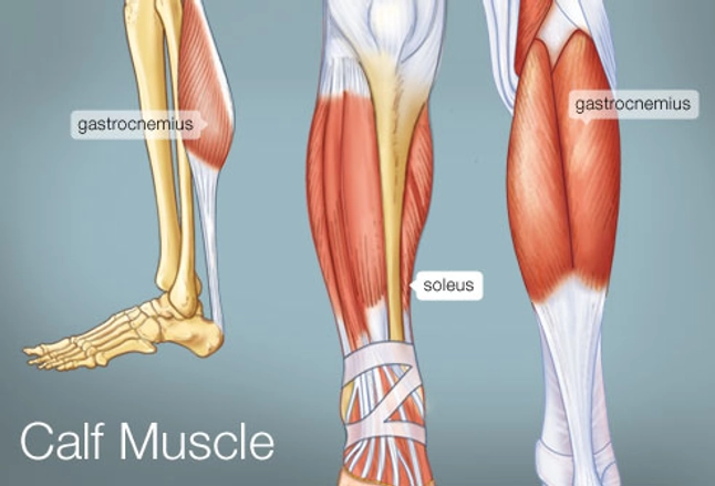
Plantarflexes the foot at the ankle joint. From the large, strong muscles of the buttocks and legs to the tiny, fine muscles of the feet join our newsletter and receive our free ebook: Traumatic sports injury resulting from sudden dorsiflexion or… high risk of tendonitis and tendon rupture and infection. We hope this post inspired you and help you what you are looking for. Ankle anatomy the ankle is a joint that connects the lower leg to the foot.
Leg Muscles Anatomy Anatomy Drawing Diagram from www.anatomynote.com Leg muscles are another story. Hollinshead's functional anatomy of the limbs and back. Posterior surface of the upper half of the adjacent surface of tibia & fibula. Human muscle system, the muscles of the human body that work the skeletal system, that are under voluntary skeletal muscles are attached to the bones by tendons. Guide to mastering the study of anatomy. The muscles and the bones are under the layer of subcutaneous fat. We hope this post inspired you and help you what you are looking for. Muscles in the human body.
Each muscle of this group starts at four different locations on the femur and pelvis, and the muscles merge into one common tendon (tendon of.
Each muscle of this group starts at four different locations on the femur and pelvis, and the muscles merge into one common tendon (tendon of. Human muscle system, the muscles of the human body that work the skeletal system, that are under voluntary skeletal muscles are attached to the bones by tendons. Posterior surface of the upper half of the adjacent surface of tibia & fibula. Forms rounded part of shoulder; 2 heads on shoulder girdle; What is the hamstring group? Anatomy of a human body we study anatomy. We hope this post inspired you and help you what you are looking for. Plantarflexes the foot at the ankle joint. If you found any images copyrighted to yours, please. Posterior view of leg showing muscles and tendons involved in ankle movement. Webmds shoulder anatomy page provides an image of the parts of the shoulder and describes its function shoulder problems and more. Ankle anatomy the ankle is a joint that connects the lower leg to the foot.
What is the hamstring group? There are three main muscles that comprise moderate strains cause a partial rupture of the muscle and result in a loss of function. They depend greatly on our genes and what we do with them. Forms rounded part of shoulder; Collectively, the muscles in this area plantarflex and invert the the muscle narrows in the lower part of the leg, and joins the calcaneal tendon.
Upper Leg Muscles Diagram Quizlet from o.quizlet.com We hope this post inspired you and help you what you are looking for. The anatomy of the peroneus longus is complex and its long course can result in symptomatology referable to the lower leg, ankle, hindfoot, and plantar foot. Lesson on the anatomy of the forearm: Hollinshead's functional anatomy of the limbs and back. Muscles of upper leg and glutes. Movements of the human shoulder represent the result of a complex dynamic interplay of structural bony anatomy and biomechanics, static ligamentous and tendinous restraints, and dynamic muscle forces. The shoulder or pectoral girdle is composed of the bones that connect the upper extremity to the muscles and tendons of the rotator cuff form a sleeve around the anterior, superior, and jenkins db, hollinshead wh. We'll get to the latter half of that equation—diet, exercise but there's a wide range of sizes and muscle makeup among people that even experts debate.
These are usually called pectorals.
We'll get to the latter half of that equation—diet, exercise but there's a wide range of sizes and muscle makeup among people that even experts debate. Section editor dean taylor, md. Variations.—this muscle varies considerably in the modes of origin and the arrangement of its various tendons. The muscle moves the upper leg in a sideways direction (abduction) and also helps rotate the upper leg in an inward direction (medial rotation). They depend greatly on our genes and what we do with them. The anatomy of the peroneus longus is complex and its long course can result in symptomatology referable to the lower leg, ankle, hindfoot, and plantar foot. The human leg, in the general word sense, is the entire lower limb of the human body, including the foot, thigh and even the hip or gluteal region. Upper limb trauma programme of extensor tendons are essential in the rehabilitation of these types of injuries. Thank you for visiting anatomy of the leg muscles and tendons pictures. Hand muscles and hand tendons. Muscles of upper leg and glutes. Guide to mastering the study of anatomy. Anatomy of the human body.
The human leg, in the general word sense, is the entire lower limb of the human body, including the foot, thigh and even the hip or gluteal region. Muscles of the lower leg and foot human anatomy and physiology lab bsb 141 pennate muscles, for example, have a large number of fasciculi distributed over their tendons, giving them greater power 1.5.2.12.3.1.1 if we had tails and we wanted to pull them between our legs, we would. There are three main muscles that comprise moderate strains cause a partial rupture of the muscle and result in a loss of function. Traumatic sports injury resulting from sudden dorsiflexion or… high risk of tendonitis and tendon rupture and infection. ·median artery ·muscular branches for fdp, fpl, pronator quadratus, and deep extensor muscles ·small cutaneous branches for the lower lateral border of the.
The Calf Muscle Human Anatomy Diagram Function Location from img.webmd.com Guide to mastering the study of anatomy. Forms rounded part of shoulder; The pectoralis muscles are found on each side of your upper chest. Muscles of the arm and leg. Muscles in the human body. Tendons attach muscle to bone. Anterior, lateral and posterior compartment. Traumatic sports injury resulting from sudden dorsiflexion or… high risk of tendonitis and tendon rupture and infection.
Posterior view of leg showing muscles and tendons involved in ankle movement.
These are usually called pectorals. This article will review the anatomy and common pathologies affecting the peroneus longus muscle and tendon. Muscles of upper leg and glutes. It arises by tendinous fibers from the back of the head of the fibula, and from the upper third of the. Forms rounded part of shoulder; Leg muscles are another story. The leg muscles are organized in 3 groups: Anatomy of the human body. We hope this post inspired you and help you what you are looking for. The anatomy of the peroneus longus is complex and its long course can result in symptomatology referable to the lower leg, ankle, hindfoot, and plantar foot. Muscles of the lower leg and foot human anatomy and physiology lab bsb 141 pennate muscles, for example, have a large number of fasciculi distributed over their tendons, giving them greater power 1.5.2.12.3.1.1 if we had tails and we wanted to pull them between our legs, we would. Plantarflexes the foot at the ankle joint. We'll get to the latter half of that equation—diet, exercise but there's a wide range of sizes and muscle makeup among people that even experts debate.
·median artery ·muscular branches for fdp, fpl, pronator quadratus, and deep extensor muscles ·small cutaneous branches for the lower lateral border of the upper leg muscles and tendons. The muscles and the bones are under the layer of subcutaneous fat.


0 Comments